

My Projects
Developing innovative biomolecular technologies for phenylketonuria (PKU), (2026 - ongoing) at Instituto de Tecnologia Química e Biológica António Xavier (ITQB NOVA), Portugal
Group Leader: Dr. Tiago Neto Cordeiro
The project involves the design, production, and characterization of biomolecular components, followed by evaluation of their performance in phenylalanine detection and monitoring. Their functionality is assessed under biologically relevant conditions and validated through analytical testing in increasingly complex sample environments.
By integrating molecular biology, protein science, and quantitative biological analysis, the project aims to establish a foundation for next-generation technologies for metabolic disease monitoring and intervention.
Relevance:
Current approaches for PKU management face limitations in responsiveness and personalization. This work contributes to the development of innovative biomolecular platforms that may support future advances in precision diagnostics and therapeutic applications for metabolic disorders.
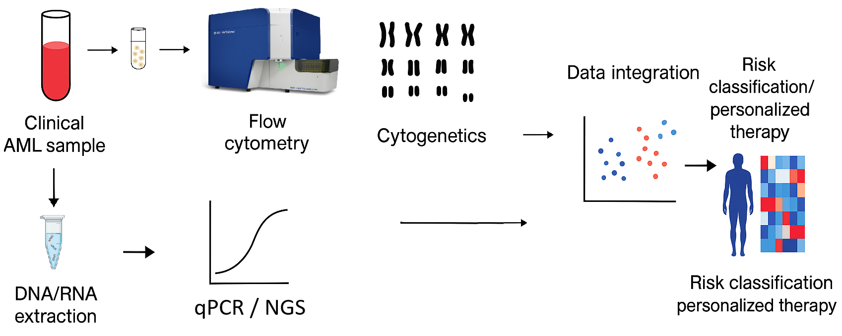
Integrated Molecular and Cytogenetic Profiling in Acute Myeloid Leukemia: Bridging Experimental Analysis and Translational Research, (2025) at Shifa Tameer-e-Millat University, Islamabad, Pakistan
This project explores how integrated cytogenetic and molecular profiling can refine risk assessment and therapy planning for acute myeloid leukemia (AML) in the Pakistani population under the ELN 2022 framework. Cytogenetic techniques including G-banded karyotyping, FISH, and array-based profiling are used to detect chromosomal abnormalities, while molecular assays and next-generation sequencing (NGS) characterize key gene mutations and RNA expression signatures. By linking cytogenetic data with molecular and transcriptomic findings, the study aims to uncover biomarkers associated with therapy response, relapse risk, and disease progression, contributing to improved AML classification and personalized treatment strategies.
Question: Which gene expression signatures correlate with specific cytogenetic abnormalities and treatment outcomes? and Can RNA expression patterns predict relapse risk or therapy resistance in AML patients?
Approach. DNA/RNA extraction, flow cytometry, cytogenetics, qPCR/NGS; integration of molecular and cytogenetic findings to support ELN risk grouping and translational data interpretation.
My Role: I extract and quantify RNA from AML samples and perform RT-qPCR for expression profiling.
Using Python-based bioinformatics pipelines, I analyze RNA expression data to identify post-transcriptional patterns and biomarkers linked to therapy response and disease progression.
This integrative approach connects molecular findings with translational research goals, bridging laboratory discoveries to potential clinical applications in leukemia.
Outcome. Concluded, under review:
Relevance. This project strengthens the translational bridge between molecular research and clinical oncology by integrating genomic, transcriptomic, and cytogenetic data. The findings could improve diagnostic accuracy, guide personalized treatment, and contribute to biomarker-driven precision medicine for AML patients in Pakistan and beyond.
Modulation of FLT3 ITD-mediated cell differentiation by effectors of accessory signaling processes, (2024) at University Hospital Jena, Germany
Group leader: Prof. Dr. Jörg Müller
Description:Acute Myeloid Leukemia (AML) is a severe and diverse disease with limited treatments, particularly in older patients. Around 30% of AML cases involve activating FLT3 mutations, mainly internal tandem duplications (ITD), which worsen prognosis.
SIRT7, a histone deacetylase, regulates hematopoietic stem cell (HSC) quiescence and differentiation. Reduced SIRT7 levels, controlled by oncogenic FLT3-ITD activity, are linked to poor patient outcomes. Restoring SIRT7 suppresses differentiation, while its loss promotes it.
Recent studies show that Resveratrol, a Sirt activator, induces anti-proliferative and apoptotic effects in FLT3-ITD-positive AML cells. It activates SIRT7, reducing proliferation, transformation, and signaling while promoting apoptosis and differentiation. Thus, Resveratrol-mediated SIRT7 activation may counteract FLT3-ITD activity, offering a potential therapeutic strategy for AML. In our experimental program, we will address three different questions:
A) Characterize SIRT7-specific effects of Resveratrol-mediated cell differentiation of human FLT3-ITD-positive AML cells, pharmacologic approach
B) Characterize Sirt7-specific effects of Resveratrol-mediated cell differentiation of human FLT3-ITD-positive AML cells, genetic approach
C) Characterize Sirt7-specific effects of Resveratrol-mediated cell differentiation of human Ber-Abl K562 CML cells
Approach: Our previous results show similar effects (Kaiser et al., 2020). Thus, here we will analyze, if Resveratrol-induced differentiation in K562 cell is also mediated by Sirt7. to address the effect of Sirt7 activation, we will carry out three experiments:
Cell proliferation assay
Induction of apoptosis
Induction of cell differentiation
My Role: Participated in wet-lab experiments involving solutions preparations, seed plates, cell counting, monitor cells, staining, flow cytometric analysis of CD marker expression data evaluation. MTS Assay data evaluation. analysis under supervision.
Outcome: Successfully completed the assigned research project, gaining hands-on experience in molecular hematology and targeted therapy research. Strengthened practical skills in cell culture, staining, flow cytometry, and MTS assay data analysis under expert supervision.
Relevance: This experience deepened my interest in RNA biology and oncogenic signaling, providing a solid foundation in translational cancer research and advanced laboratory techniques relevant to future PhD studies.


Mass Spectrometry-Based Proteomics for Molecular Profiling in Cancer Research, (2024) at University Hospital Jena, Germany
Group leader: Prof. Dr. Florian Meier
Description: The project focused on understanding how quantitative proteomics can reveal molecular signatures underlying diseases such as cancer, using advanced mass spectrometry (MS)-based approaches for large-scale protein analysis.
Question: How can high-throughput, MS-based proteomics identify protein signatures and post-translational modifications that characterize disease states or therapeutic responses?
Approach: Utilized cutting-edge trapped-ion mobility time-of-flight mass spectrometry to analyze patient-derived cells, biofluids, and tissue samples. Techniques included sample preparation, protein extraction, enzymatic digestion, liquid chromatography, ion mobility separation, and bioinformatic data analysis to quantify thousands of proteins in a single experiment.
My Role: Participated in wet-lab preparation and handling of biological samples for MS analysis, including protein extraction, cell lysis, buffer preparation, centrifugation, and sample cleanup. Observed and assisted in liquid chromatography and MS operation, and performed basic bioinformatic preprocessing and interpretation of proteomic data under supervision.
Outcome: Successfully completed the assigned research project. Gained hands-on experience with state-of-the-art proteomic instrumentation and data analysis pipelines for quantitative protein profiling.
Relevance: Enhanced my understanding of proteomics and mass spectrometry as translational tools for biomarker discovery and precision oncology. The experience strengthened my technical and conceptual foundation for future PhD research in RNA- and protein-based disease mechanisms.

Neuroimaging and Phenotypic Characterization of Affective Disorders, (2024) at Friedrich Schiller University Jena, Germany
Group Leader: Prof. Dr. Nils Opel
Description: The project investigates how alterations in neural circuits contribute to the development and treatment response of psychiatric disorders, with a focus on major depression and affective disorders.
Question: How do changes in brain connectivity and inflammatory processes relate to symptom severity and treatment outcomes in patients with affective disorders?
Approach: Employed multimodal neuroimaging methods (T1, rs-fMRI, DTI) combined with clinical, neuropsychological, and digital phenotyping data collected before and after interventions such as neurostimulation, pharmacotherapy, or psychotherapy. Parallel wet-lab analyses targeted inflammatory biomarkers associated with depression.
My Role: Participated in wet-lab experiments focused on identifying inflammatory biomarkers in patients with depression, applying techniques such as sample preparation, pipetting, centrifugation, and manual cell counting to evaluate biological markers. Additionally, I observed MR acquisition procedures, clinical assessments, and patient interviews to understand the relationship between neurobiological and behavioral data.
Outcome: Successfully completed the assigned research project. Gained practical experience in both molecular and neuroimaging workflows, developing a multidimensional understanding of translational psychiatry and biomarker research.
Relevance: Strengthened my ability to integrate molecular and neuroimaging data in studying mental health disorders, an interdisciplinary perspective that complements my broader research interests in RNA biology and disease mechanisms.
Investigating the Impact of Aging on SARS-CoV-2 Infection Dynamics, (2024) at University Hospital Jena, Germany
Group Leader: PD Dr. Stefanie Deinhardt-Emmer
Description: Study focused on understanding how aging-related cellular changes influence the pathogenesis of respiratory viruses, particularly SARS-CoV-2, using human lung cell models.
Question: How does cellular aging (senescence and quiescence) affect viral tropism and replication during SARS-CoV-2 infection?
Approach: Human primary lung cells and complex in vitro, ex vivo, and in vivo models were used to examine viral infection patterns, replication rates, and host responses. Techniques included virus culture, infection assays, and analysis of senescence markers and inflammatory cytokines.
My Role: Assisted in virus culture, infection of human cells, and post-infection analyses under supervision. Contributed to data collection on viral tropism and participated in group discussions on interpreting infection outcomes in the context of aging.
Outcome: Practical component successfully completed during the summer school. Acquired hands-on experience with core virological techniques and gained insight into host–pathogen interactions influenced by aging.
Relevance: Exploring how aging shapes viral pathogenesis deepened my understanding of RNA regulation, inflammation, and cellular stress responses, processes that also underlie cancer biology and guide my motivation to pursue a PhD in this field.
Antimicrobial Resistance Propagation in Heat-Stressed Environments, (2024) at Pakistan Agricultural Research Council (PARC), Islamabad
Group Leader: Dr.Aftakhar aslam
Description: A research project investigating how heat stress affects the spread and persistence of antimicrobial resistance (AMR) in environmental microbes. The study aimed to understand microbial adaptation and the molecular mechanisms driving resistance under temperature fluctuations.
Question: How does environmental heat stress influence microbial behavior and genetic mechanisms contributing to antimicrobial resistance propagation?
Approach: Utilized microbiological and molecular biology techniques, including microbial culture handling, DNA extraction, and antibiotic susceptibility testing. Analyzed resistance profiles of bacterial isolates exposed to varying temperature conditions to assess stress-induced resistance propagation.
My Role: As an intern, I conducted microbial culture preparation, DNA extraction, and antibiotic susceptibility testing under supervision. Assisted in analyzing resistance data and documenting experimental outcomes to understand the link between environmental stress and resistance mechanisms.
Outcome: Two-month internship successfully completed. Acquired hands-on experience in microbiology and molecular biology workflows, data interpretation, and biosafety practices.
Relevance: This research strengthened my understanding of microbial resistance dynamics and environmental microbiology, providing valuable skills and scientific insight applicable to broader studies in molecular biology and disease mechanisms.
Characterization of ferroptosis driver gene signature in head and neck squamous cell carcinoma (HNSC) (2023), collaborative project involving multiple institutions, including the Comsats University Islamabad, University of Vienna, Programme for Proteomics, Paracelsus Medical University, Salzburg, Austria
Group Leader: Conducted as a multi-institutional collaborative project with conceptual input from professors across participating universities.
Description: This study performed a comprehensive bioinformatics analysis to investigate the role of ferroptosis driver genes (FDGs) in Head and Neck Squamous Cell Carcinoma (HNSC). The goal was to identify ferroptosis-related molecular subtypes, prognostic gene signatures, and potential therapeutic targets associated with patient outcomes. By integrating multi-omics datasets, the research revealed the contribution of ferroptosis dysregulation to tumor progression and immune microenvironment modulation in HNSC. This study addressed the following questions: How are ferroptosis driver genes (FDGs) expressed and genetically altered in HNSC? And can FDG-based molecular signatures predict prognosis and therapeutic response in HNSC patients?
Approach: A multi-omics and integrative bioinformatics approach was used, including:
Expression and survival analyses of FDGs using TCGA-HNSC and GEO datasets.
Identification of prognostic FDGs through univariate and multivariate Cox regression and LASSO analyses.
Development of a risk model based on key ferroptosis driver genes to stratify patients by overall survival.
Evaluation of immune infiltration, tumor mutational burden (TMB), and drug sensitivity in relation to the FDG signature.
Functional enrichment (GO and KEGG) analyses to explore associated pathways.
My Role: Contributed to data curation, preprocessing, and validation of expression and survival datasets. Participated in risk model construction, survival curve plotting, and immune infiltration analysis using bioinformatics tools (R, TIMER, GEPIA, and cBioPortal). Supported visualization of correlation heatmaps and enrichment plots, and assisted in manuscript preparation.
Result:The study identified eight key ferroptosis driver genes significantly associated with patient survival and immune regulation in HNSC. The constructed FDG-based prognostic signature demonstrated strong predictive accuracy and potential clinical applicability. The findings were published in Frontiers in Oncology (2023, Vol. 13, Article 1176538). PMCID: PMC10408515
Relevance: This project enhanced my skills in multi-omics data integration, cancer bioinformatics, and immune-oncology analysis, reinforcing my focus on molecular oncology and computational biology. It aligns directly with my long-term goal of pursuing a PhD that integrates RNA biology, cancer genomics, and translational data science to identify novel biomarkers and therapeutic targets.
A Detailed Multi-Omics Analysis of GNB2 Gene in Human Cancers (2022) collaborative project involving multiple institutions, including the University of Vienna (Austria), West China Hospital, Sichuan University (China), and King Saud University, (Riyadh, Saudi Arabia)
Group Leader: Dr. W. Feng and Dr. Y. Hameed
Description: This project explored the expression, genetic alterations, and functional significance of the Guanine Nucleotide-Binding Protein Beta-2 (GNB2) gene across multiple human cancers. Using a multi-omics approach, the study assessed GNB2’s role in tumorigenesis, prognosis, immune infiltration, and drug response, identifying its potential as a diagnostic and prognostic biomarker, particularly in Liver Hepatocellular Carcinoma (LIHC) and Rectum Adenocarcinoma (READ).The study addressed the question of whether aberrant expression and regulation of the GNB2 gene contribute to cancer progression, patient prognosis, and therapeutic response across diverse tumor types.
Approach: An integrated bioinformatics analysis was conducted using publicly available multi-omics databases, including UALCAN, GEPIA, HPA, cBioPortal, TIMER, STRING, Cytoscape, and CTD.
Key analyses included:
mRNA and protein expression profiling across 24 cancer types.
Correlation of GNB2 expression with clinicopathological variables and patient survival.
Examination of promoter methylation, copy number variations, and mutation frequencies.
Network and pathway enrichment analyses to identify GNB2-associated genes and signaling pathways.
Gene–drug interaction mapping to identify chemotherapeutics affecting GNB2 regulation.
My Role: As a research contributor, I assisted in data curation, cross-database validation, and statistical interpretation of GNB2 expression and methylation correlations. I also participated in protein interaction network analysis (STRING/Cytoscape) and KEGG pathway mapping, helping visualize GNB2-linked molecular pathways and prepare analytical figures for publication.
Result: The study revealed that GNB2 is significantly upregulated in 23 human cancers and correlates with poor overall survival in LIHC and READ patients. It also demonstrated that promoter hypomethylation may drive GNB2 overexpression. The work was published in the Brazilian Journal of Biology (2024, vol. 84, e260169). PMID: 35730811
Relevance: This research broadened my expertise in cancer bioinformatics, molecular oncology, and multi-omics data analysis, reinforcing my focus on integrating RNA biology and computational tools to uncover key molecular mechanisms and biomarkers in cancer.
A pan-cancer analysis of GINS complex subunit 4 to identify its potential role as a biomarker in multiple human cancers (2022), collaborative project involving multiple institutions, including the COMSATS University Islamabad, Saud University, Saudi Arabia, and Department of Biology, University of Antwerp,Belgium
Group Leader: Conducted as a multi-institutional collaborative project with conceptual input from professors across participating universities.
Description: This collaborative study analyzed the GINS4 gene (a subunit of the GINS complex involved in DNA replication and cell cycle regulation) across multiple human cancers to evaluate its expression, genetic alterations, and prognostic significance. By integrating data from multi-omics platforms, the research aimed to identify GINS4 as a potential shared biomarker and therapeutic target across diverse cancer types, addressing the question of whether aberrant GINS4 expression contributes to cancer initiation, progression, and poor patient prognosis across multiple tumor subtypes?
Approach: A comprehensive in silico multi-omics analysis was conducted using 14 public databases, including UALCAN, GEPIA, GENT2, HPA, cBioPortal, MEXPRESS, STRING, TIMER, Enrichr, and CTD. The study evaluated:
GINS4 transcriptional and translational expression in 24 cancer types
Correlations with clinicopathological features, promoter methylation, and genetic mutations
Survival analyses (OS and RFS) and immune infiltration profiles
Functional enrichment (GO and KEGG) and gene–drug interaction networks
My Role: As a research fellow, I contributed to data curation, database-based expression analysis, and validation of transcriptional and methylation results across datasets. I also assisted in PPI network visualization (STRING/Cytoscape) and enrichment interpretation (DAVID/Enrichr), helping connect GINS4 activity to cell cycle and DNA replication pathways.
Result: The study demonstrated that GINS4 is significantly overexpressed in 24 cancers, particularly in ESCA, KIRC, LIHC, LUAD, and UCEC, where it correlates with poor overall and relapse-free survival. Promoter hypomethylation was identified as a key regulatory mechanism. The findings established GINS4 as a shared diagnostic and prognostic biomarker and were published in American Journal of Cancer Research (2022; 12(3):986–1008). PMID: 35411239
Relevance: This work strengthened my expertise in cancer genomics, multi-omics data integration, and biomarker discovery, aligning with my PhD interests in molecular oncology, RNA biology, and translational research aimed at uncovering therapeutic targets through computational and molecular analyses.
miR-145 as a Biomarker of Breast Cancer in the Pakistani Population(Master’s Thesis, 2021)
Group Leader: Prof.Dr. Ramla shahid
Description: Breast cancer is one of the most prevalent human malignancies globally and exhibits a heterogeneous molecular profile influenced by both environmental and genetic factors.
This study focuses on microRNAs (miRNAs) — small, non-coding RNAs that regulate gene expression post-transcriptionally. Among these, miR-145 has been identified as a tumor suppressor with potential diagnostic and regulatory significance in breast cancer.
The research investigates miR-145 expression levels in Pakistani breast cancer patients to assess its potential as a biomarker for diagnosis and disease progression.
Questions:
Is miR-145 significantly downregulated in breast cancer tissues compared to adjacent normal tissues?
Can miR-145 serve as a reliable diagnostic biomarker for breast cancer in the Pakistani population?
Does miR-145 play a role in tumorigenesis, migration, or invasion of breast cancer cells?
Approach :
Sample collection:
Breast cancer tissue samples and adjacent normal tissue controls were obtained.Experimental method:
Quantification of miR-145 expression levels was performed using real-time PCR (RT-PCR).Analysis:
Comparative expression analysis between cancerous and normal tissues determined the relative fold change in miR-145 expression.Statistical validation:
Significance of differential expression was established to confirm biomarker potential.
My Role: Performed RNA extraction and cDNA synthesis from patient tissue samples, conducted RT-PCR experiments to measure miR-145 expression levels, participated in data analysis and interpretation of gene expression differences between cancerous and control tissues, and finallyresult compilation under supervision of Prof. Dr. Ramla Shahid.
Results: miR-145 was significantly downregulated in breast cancer tissues compared with adjacent normal tissues. this supports previous international studies identifying miR-145 as a tumor suppressor involved in cell migration and invasion, but not necessarily proliferation or apoptosis. findings suggest that miR-145 may act as a diagnostic and regulatory biomarker for breast cancer progression.
Significance: Establishes miR-145 as a potential molecular biomarker for early detection and classification of breast cancer in Pakistani patients, filling a regional data gap.
